Stretching – Proprioceptiv Neuromuscular Facilitation (PNF)

Proprioceptive neuromuscular facilitation (PNF) was first developed by Herman Kabat MD, Margaret Knott PT, in the 1940’s to treat neurological dysfunctions. PNF stretching is currently the fastest and most effective way known to increase static-passive flexibility. PNF stretching is currently the fastest and most effective way known to increase static-passive flexibility.

PNF refers to any of several post-isometric relaxation stretching techniques in which a muscle group is passively stretched, then contracts isometrically against resistance while in the stretched position, and then is passively stretched again through the resulting increased range of motion.

Indications of PNF

  • Loss of range of motion.
  • Muscle tightness.
  • Muscle cramp.
  • Loss of flexibility.

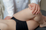
In PNF stretching, a partner is required. The partner gently moves a limb into position, usually moving it just into a place where the person feels some discomfort, but no pain. The limb can simply be held in place for a set period for a passive stretch, or the person stretching can be asked to contract the muscles against the partner’s hand for a resistance stretch. After the stretch, range of motion is usually slightly increased, and when this is done on a regular basis, range of motion overall will improve.

It is important to avoid overuse of PNF stretching, because people can be injured if their muscles are stretched too far, too soon.

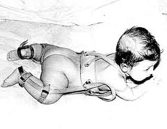
Congenital dislocation of the hip

The hip joint includes the articulation of the femoral head (of femur) and the acetabulum of the pelvis. Dislocation occurs when the ball–shaped head of the femur comes out of the cup–shaped acetabulum. Congenital hip dislocation is much more common in girls than boys.

Risk factors:

  • Children with a family history of hip dysplasia
  • Babies born in breech position

Examination:

The criteria for the diagnosis of congenital dislocation of the hip include both physical and radiographic findings. Dynamic ultrasound is used to assess hip stability and acetabular development in infants and babies under 5 monts. An x-ray of the hip joint may help diagnose the condition in older infants and children.

The Ortolani– and Barlow tests (physical examinations) are the most common clinical tests for newborn babies. These tests are useful in neonates but become difficult by 2-3 months of age.

Physical signs for late dislocation :

  • one leg appears shorter than the other
  • include asymmetry of the gluteal thigh or labral skin folds,
  • buttock flattening,
  • asymmetrical thigh skin folds,
  • decreased abduction on the affected side,
  • standing or walking with external rotation of the affected leg.
  • limited abduction when fully flexed.
  • older children may walk limping

Treatment

The principal treatment for Congenital dislocation of hte hip is conservative, especially if diagnosed early. Early diagnosis and treatment is important for a good outcome. In a newborn infant with a good reduction, there is a very good chance of full recovery. The success of treatment depends on the age of the child, and the adequacy of the reduction.

The most important treatment are:

  • Physical therapy, it is important to to improve treatment results and to decrease the risk of complications.
  • A dynamic flexion-abduction orthosis – Pavlik-harness – left in place at all times, is used to maintain hip reduction. The treatment must be continued until the hip is stable and xrays or ultrasound examinations are normal, but a Pavlik-harness is contra-indicated in children older than 6 months and when the hip is irreducible.

Photo: Sulypont Pain and Rehabilitation Clinic

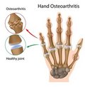
    What Is Osteoarthritis?

    Osteoarthritis is the most common form of arthritis. Osteoarthritis occurs when the protective cartilage on the ends of your bones wears down over time.

    The most affected joins, are: hands, neck, lower back, knees and hips. Osteoarthritis affects just joints, not internal organs.

    Risk factors :

    • Being overweight (extra weight causes more wear and tear)
    • Getting older (usually appear in middle age)
    • Fractures or other joint injuries
    • Certain occupations
    • Genetic factors
    • Other diseases which change cartilage structure(metabolic or hormonal disorders)
    • Your sex – Women are more likely to develop osteoarthritis than men.

    Signs and symptos:

    • Pain in the affected joint(s), that usually geting worse later, after time causing pain even at rest or limited motion.
    • Stiffnes, swelling, warmth, and creaking (crepitus) of the affected joints.
    • The ligaments and muscles around the joint become weaker and stiffer
    • Joint tenderness
    • Local inflammation
    • Limited range-of-motion
    • Muscle weakness
    • Complete loss of the cartilage cushion causes friction between bones
    • Limping ( knee- or hip degeneration)
    • Progressive cartilage degeneration of the knee joints can lead to deformity and outward curvature of the knees,
    • Deformation of the small joints of the fingers and the toes
    • Heberden’s node, osteophyte formation

    About treatment:

    Treatments include physical therapy, medicines and sometimes surgery.

    The goal of treatment is :

    • to reduce joint pain
    • to reduce inflammation
    • improving and maintaining joint function with physical therapy.
    • sometimes, the pain, limping, and joint dysfunction may not respond to medications and physical therapy, they need total knee replacement (surgical procedures) Osteoarthritis is the most common reason for knee replacement
    • Physical therapy is an essential part of rehabilitation after total knee replacement.
    • Weight loss if patient is overweight

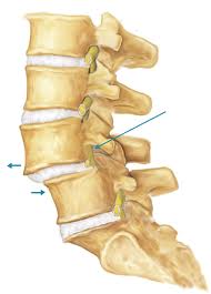
    Spondylolisthesis

    Spondylolisthesis is a condition in which a bone (vertebra) in the spine slips out of the proper position onto the bone below it. Forward slippage of one vertebra on another is referred to as anterolisthesis, while backward slippage is referred to as retrolisthesis. Though it can occur at the level of cervical vertebrae, it occurs most often at the level of the lumbar vertebrae. Spondylolisthesis usually occurs between the fifth bone in the lower back (lumbar vertebra) and the first bone in the sacrum (pelvis) area (L5-S1)

    Major types of lumbar spondylolisthesis:

    • Congenital spondylolisthsis
    • Traumatic spondylolisthsis
    • Degenerative spondylolisthesis
    • Pathologic spondylolisthesis

    What are the symptoms of spondylolisthesis? (more…)

    Perthes disease, Legg-Calve-Perthes disease

    A child who has a hip problem may feel pain in the hip, groin, thigh, or knee. A child in pain may limp or be unable to stand, walk, or move the affected leg.

    Legg-Calvé-Perthes disease is a childhood condition that affects the hip, is a temporary hip condition in which the femoral head loses its blood supply (avascular necrosis). Perthes disease usually involves just one hip, but both hips can be affected. It is rare for a child to have whole femur-head involvement.

    The four stage of Perthes disease:

    • femoral head becomes more dense with possible fracture of supporting bone
    • fregmentation and reabsorption of bone
    • reossification
    • healing (new bone reshapes) (more…)

    Osgood-Schlatter syndrome

    Osgood-Schlatter syndrome it’s really not a disease, but an overuse injury of the knee. This is one of the most common causes of knee pain in adolescents.

    The condition affects guys and girls, (boys ages 13 to 14 and girls ages 11 to 12) especially who are active in sports or participating in competitive sports, involving deep knee bends, jumping, and running, and swift changes of direction. (soccer, basketball, skating, volleyball, etc.) Usually only one knee is affected, but both can be.

    Osgood-Schlatter syndrome is mainly diagnosed by:

    • physical examination
    • X-ray
    • Ultrasound scan.
    • MRI (magnetic resonance imaging)

    Risc factors:

    • Age between 11 and 15 years
    • Male sex
    • Rapid skeletal growth
    • Repetitive jumping sports
    • Tight quadriceps (front thigh) muscles,
    • Tight hamstrings (back thigh) muscles (more…)

    The human spine

    The human spine is a marvel of engineering. The spinal column has a pyramidal design with the largest vertebrae at the base. Composed of 24 tightly connected vertebrae, each vertebrae offers 4 degrees of movement from the front to the back, providing a column that is both very strong and flexible.

    The spinal column is literally a column that rests on the pelvis as its base of support and is kept perpendicular by the cable-like functioning of the surrounding musculature. Should this musculature cause the vertebrae to curve, tilt and/or twist then the stage is set for a host of back problems.

    For example, if the surrounding musculature causes a vertebrae to tilt just 4 degree from its proper position, the lower vertebrae experiences a 30% weight increase on the loaded side. Backaches, pinched nerves, herniated discs, etc. are all a result of such vertebral misalignments. Most cases of lumbago and sciatica are due to such pinched nerves emanating from between improperly aligned vertebrae.

    Since the spinal column is dependant on the pelvis for its base of support, practically all spinal deviations are really an extension of pelvic deviations. By restoring core structural stability to the pelvis and symmetrical functioning the surrounding musculature, most spinal deviations the problems of the lower, middle, and upper back tend to resolve themselves nicely within a matter of months.

    About Toes

    Due to constant enclosure within shoes as well as the proliferance of smooth, flat surfaces, most people hardly use their toes anymore. Toes do have a number of important responsibilities to fulfill, however. They serve as messengers to the brain, for example, constantly sending reports about the terrain under them “on the ground”. They also assist in fine tuning the balance and support of the body.

    Despite their small size, toes can be a great source of discomfort if structural deviations should start to develop anywhere throughout the body. (more…)

    About Elbow and Fingers

    Fingers Wrists Wrists Elbows Elbows Neck and head Shoulders Neck and head Shoulders Spine Spine Spine Spine Spine Hips Hips Hips Hips Knee Toes Feet Feet Knee Knee Knee Feet Feet Toes

    Movement of the elbow is mainly controlled from above by the shoulder. Should the responsible postural musculature no longer maintain the shoulders in proper alignment with the hips and spine, then the hinge joint functioning of the elbow will be called upon to compensate for a dysfunctional ball-and-socket joint of the shoulder. Such impossible demands placed upon the elbow joint soon lead to increased strain and wear and result in a sensitive and painful elbow joint.

    Most elbow problems, therefore, find their origin in misalignments occurring at the shoulder and in practice practically all elbow problems can be lastingly resolved by restoring proper postural alignment across the shoulder girdle and then down through the arm.The pelvis serves as the “foundation” of the body’s skeletal structure. (more…)

    About shoulders

    Of all the joints of the body, the shoulder joint is the most flexible. This great range of motion is achieved by mounting a very flexible ball-and-socket joint on a stable, yet very mobile platform consisting of the shoulder blade and collar bone. Thus the entire shoulder girdle can be positioned up, down, back, and forth as needed to provide the best positioning from which the very flexible ball-and-socket joint can function. Extended periods of sitting or asymmetric activity can result in one or both shoulders becoming fixated in a forward and/or downward position. Such shoulder deviations have become so commonplace, that forward hunching, drooping shoulders are viewed as the norm. The problem is that such compromised postures restrict the shoulder’s range of motion and force the elbows, wrists, and fingers to perform a wide array of demanding lifting, swinging, twisting functions that they were not meant to tolerate. Should repetitive motion demand be thrown onto the misaligned shoulder (tennis, typing, etc.) the compromised joint will register the condition in the form of tennis elbow, carpal tunnel syndrome or a host of other syndromes, in an attempt to quickly stop the damaging activity.

    (more…)